


[Year:2014] [Month:September-December] [Volume:8] [Number:3] [Pages:1] [Pages No:0 - 0]
DOI: 10.5005/jocgp-8-3-v | Open Access | How to cite |

[Year:2014] [Month:September-December] [Volume:8] [Number:3] [Pages:1] [Pages No:85 - 85]
DOI: 10.5005/jp-journals-10008-1167 | Open Access | How to cite |

Surgical Outcome of Ahmed Valve Implantation in Mexican Patients with Neovascular Glaucoma
[Year:2014] [Month:September-December] [Volume:8] [Number:3] [Pages:5] [Pages No:86 - 90]
Keywords: Neovascular glaucoma, Surgical outcome, Ahmed valve implantation, Glaucoma filtration surgery
DOI: 10.5005/jp-journals-10008-1168 | Open Access | How to cite |
Abstract
Purpose: To describe clinical results of Ahmed glaucoma valve implantation in Mexican patients with neovascular glaucoma (NVG). Materials and methods: We reviewed records of 60 eyes of 60 patients with NVG who underwent Ahmed valve implantation, with a follow-up period of 1 year. We identified successful and failed cases and compared baseline and follow-up characteristics to identify possible differences between both groups. Results: We classified 36 eyes (60%) as successful and 24 (40%) as failed cases. We found a significant difference in success rate in patients who had a hypertensive phase at any time during the follow-up period (OR = 5.15, CI = 1.49-20.15, p = 0.004). Patients in the success group showed a statistically significant decrease in the number of glaucoma medications 1 year after surgery (p <0.0001). We found a statistically significant difference in success rate in patients who had preoperative best corrected visual acuity (BCVA) better than logmar 0.70 (odds ratio 4.31, CI = 1.1-19.3, p = 0.03086). Conclusion: A hypertensive postoperative phase and a preoperative BCVA worse or equal to 20/100 seem to be risk factors for Ahmed valve surgical failure in patients with NVG.

[Year:2014] [Month:September-December] [Volume:8] [Number:3] [Pages:5] [Pages No:91 - 95]
Keywords: Deep sclerectomy, Intraocular pressure, Esnoper-Clip implant, AS-OCT
DOI: 10.5005/jp-journals-10008-1169 | Open Access | How to cite |
Abstract
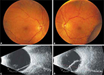
Purpose: To describe the technique of deep sclerectomy with the new Esnoper-Clip® implant, the clinical outcome and the anatomic characteristics of filtering blebs, using anterior segment optical coherence tomography (AS-OCT). Methods: A prospective case-series study was conducted in five eyes (5 patients) with open angle glaucoma. The fornix-based deep sclerectomy with Esnoper-Clip® implant was done by the same surgeon. In one case, mitomycin C was used during surgery. All participants underwent a complete ophthalmic examination and AS-OCT (Visante®) preoperatively, then at each follow-up visit, at 1 day, 1 week, 1 month, 6 months and 1 year postoperatively. Scans were obtained through sagittal and transversal plans to the implant. Results: Intraocular pressure (IOP) was significantly reduced (p < 0.05) from a mean preoperative value of 23.4 ± 8.6 mm Hg (n = 3.8 glaucoma medications) to a postoperative value of 6.0 ± 2.5 (n = 0), 10.6 ± 5.4 (n = 0), 13 ± 1.6 (n = 0.4), 12.4 ± 2.1 (n = 0.2) and 14.4 ± 1.5 (n = 0.2) at 1 day, 1 week, 1 month, 6 months and 1 year respectively. AS-OCT allowed the visualization of the two plates of the implant (scleral and suprasciliary), the trabeculodescemetic membrane and the hyporeflective spaces in the bleb wall thickness and in suprascleral and suprachoroidal localizations. An immediate postoperative hypotony and an anteriorization of the implant associated to trabeculodescemetic membrane rupture, were detected, although without significant clinical repercussions. Conclusion: Our first five deep sclerectomy with Esnoper-Clip implantation analysis suggest an effective and well-tolerated method to reduce IOP. AS-OCT is a noninvasive imaging technique that allows the anatomic analysis of the drainage mechanisms after glaucoma surgery.

[Year:2014] [Month:September-December] [Volume:8] [Number:3] [Pages:5] [Pages No:96 - 100]
Keywords: Glaucoma, Baerveldt, Tube ligation method, Absorbable ligation method, Nonabsorbable ligation method, Tube shunt surgery
DOI: 10.5005/jp-journals-10008-1170 | Open Access | How to cite |
Abstract
Objective: To investigate the early postoperative complications in two different tube ligation methods during the first 3 months in Baerveldt implant surgery. Participants: This study involved 157 eyes from 144 patients who underwent Baerveldt Implant Surgery at the Japanese Red Cross Medical Center, Tokyo, Japan. Methods: Pre- and postoperative intraocular pressure (IOP), combined surgery, postoperative time-point of tube ligation release, and postoperative complications in two different tube ligation methods [absorbable ligation method using 8-0 polyglactin suture (group A) and nonabsorbable ligation method using 7-0 nylon suture (group B)] were retrospectively reviewed. Results: After excluding eyes that had undergone combined trabeculectomy (26 eyes) and vitrectomy (2 eyes), eyes with previous tube surgery (22 eyes), and eyes that had undergone the stent method (1 eye), 30 of 28 patients in group A and 71 eyes of 71 patients in group B were found to fit the criteria of this study. The rate of successful surgical outcome was 80% in group A and 74.6% in group B (p = 0.705). During the 3 months postoperative, high IOP tended to occur more often in group B (67.6%) than in group A (46.7%) (p = 0.073), and ciliochoroidal detachment tended to occur more often in group A (10.0%) than group B (2.8%) (p = 0.154). Conclusion: The results of this study show that both ligation methods are effective, however, the selection of tube ligation method should be done in accordance with the different method-specific risks to which may occur.

[Year:2014] [Month:September-December] [Volume:8] [Number:3] [Pages:6] [Pages No:101 - 106]
Keywords: GCC, OCT, RNFL, Pre-perimetric glaucoma, Peri-metric glaucoma
DOI: 10.5005/jp-journals-10008-1171 | Open Access | How to cite |
Abstract
Purpose: To determine the importance of ganglion cell complex (GCC) analysis as a parameter for early diagnosis of glaucoma and for following glaucoma progression and to compare glaucoma progression with conventional visual field analysis using a different type of spectral-domain optical coherence tomography (SD-OCT). Materials and methods: Two hundred eyes including 68 normal eyes, 70 eyes with pre-perimetric glaucoma and 62 eyes with perimetric glaucoma were analyzed in this prospective study undertaken during Jan 2013 to Dec 2013 in a tertiary ophthalmology institute. Automated visual field examination was done to group the subjects in above three categories. The thicknesses of the GCC and retinal nerve fiber layer (pRNFL) were measured using Topcon model 2000 version 7.1 SD-OCT images and compared. The statistical analysis was carried out by z-test. Results: The average GCC was thickest in the normal group and the thickness decreased as the severity of glaucoma increased. The mean macular GCC at the start and end of the study in pre-perimetric (94.86 ± 8.31, 90.74 ± 8.46) and perimetric (82.48 ± 13.21, 79.80 ± 12.88) eyes was lower than those in normals (102.70 ± 7.19, 101.82 ± 7.42). Conclusion: Majority of the studies done on GCC analysis have used the Cirrus OCT (Zeiss). Our study has used the Topcon model 2000 version 7.1 to show that irrespective of the machine used, GCC analysis definitely plays an important role. To detect pre-perimetric glaucoma and may show progression earlier than pRNFL in pre-perimetric glaucoma.

Screening First Degree Relatives of Persons with Primary Open Angle Glaucoma in India
[Year:2014] [Month:September-December] [Volume:8] [Number:3] [Pages:6] [Pages No:107 - 112]
Keywords: Primary open angle glaucoma, Family glaucoma, Family history, Heredity history, Heredity, Risk factor, Targeted factor, Targeted screening, First degree relatives
DOI: 10.5005/jp-journals-10008-1172 | Open Access | How to cite |
Abstract
Purpose: To report the results of screening first degree relatives of persons identified with primary open angle glaucoma in a tertiary eye hospital glaucoma services. Design: A cross-sectional study of first degree relatives of persons with primary open angle glaucoma. Materials and methods: First degree relatives of patients identified with primary open angle glaucoma were invited to participate in a screening evaluation in the base hospital to detect glaucoma. All participating individuals had comprehensive eye examination including vision screening, refraction, slit-lamp biomicroscopy, applanation tonometry, gonioscopy, frequency doubling peri-metry and dilated fundus examination. Persons with definite and suspected glaucoma were subject to full threshold automated perimetry. Results: A 514 first degree relatives of 346 persons with primary open angle glaucoma, of 4972 individuals who were invited to participate attended the screening examination (Response Rate 7%). Fifty-five percent of those who attended were males and mean age of participants was 56.8 years. Sixty-eight relatives (13.3% of those screened) were detected to have definite glaucoma. Sixty percent of those detected with definite glaucoma were siblings. Fifteen percent of siblings, 4% of off-springs and 20% of parents who attended the screening examination had definite open angle glaucoma. Conclusion: Prevalence of open angle glaucoma amongst first degree relatives of persons with glaucoma is higher than in the general population as reported in previous studies. Significant barriers, however, exist in the uptake of eye care services among relatives of persons known to have primary open angle glaucoma.
Bilateral Acute Angle-closure after Intraocular Surgery
[Year:2014] [Month:September-December] [Volume:8] [Number:3] [Pages:2] [Pages No:113 - 114]
Keywords: Acetazolamide, Choroidal effusion, Surgery complication, Acute secondary angle-closure
DOI: 10.5005/jp-journals-10008-1173 | Open Access | How to cite |
Abstract
We report the case of a 75-year-old woman who developed an acute bilateral angle-closure associated with choroidal effusion a day after an uneventful cataract surgery. The same patient had undergone a similarly uneventful cataract surgery two weeks before, under the same protocol, with no postoperative complication in the other eye. Medical treatment, including the use of oral sulfamide-related drugs (acetazolamide), topical beta-blockers and steroids led to a gradual decrease in intraocular pressure (IOP) and choroidal effusion. Despite initial reports suggesting a link between sulfamide-exposure and these rare forms of angle-closure, our report would suggest a more complex pathophysiology behind this intriguing phenomenon.


© Jaypee Brothers Medical Publishers (P) LTD.

